This section describes reconstruction from serial optical sections of the eye of Lymnaea stagnalis.
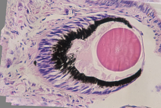
Positive color slides obtained by optical microscope of 25 serial sections of thickness 5µm were scanned via a film scanner. DeltaViewer then aligned the scanned images; the above is one of them.
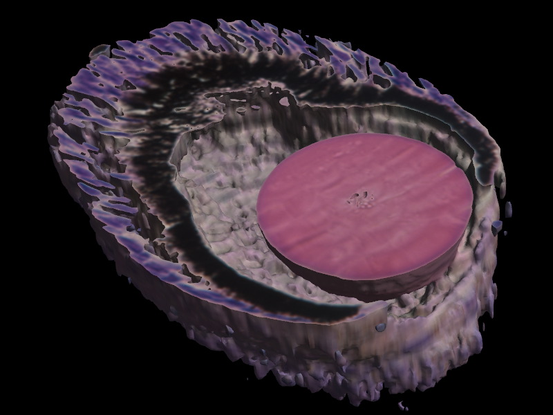
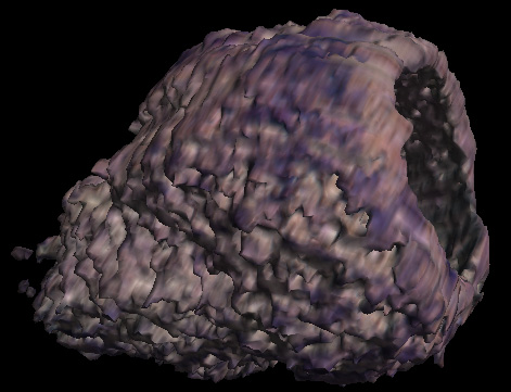
A separate image of micrometer taken with the same magnification setup reveals that 50µm corresponds to 130 pixels on the image. Thus, the 5µm gap between the slices is equivalent to 13 pixels. To correct scale in depth, we use Scale Images menu and set z-scale=13. The screenshot at the top of this page was created this way. We can change selection and reconstruct only the pigment layer (the dark part in the image), if we want:
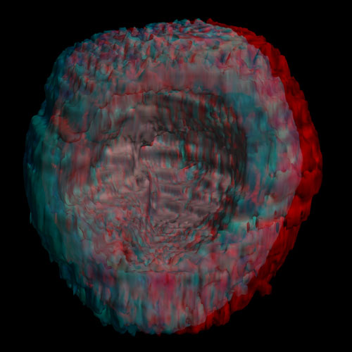
Anaglyph of the pigment layer clearly shows the irregular shape of the bottom part of its inside.