The sample image files obtained from Ganglion.sit.bin provided in the download section below are from Leica confocal laser microscope via TCS NT software. As you decompress the file, you get a folder named Ganglion containing two sub-folders lamp-HTG3-HT and lamp-HTG3-G each containing 121 TIFF files of red and green components respectively (together with some log files which are irrelevant to the operation).
To read these files in DeltaViewer, you choose Read Files(Red) menu. In the "Choose File" dialog that appears, go to the lamp-HTG3-HT folder and choose the first file c02f0001.TIF. DeltaViewer then automatically reads in the files c02f0002.TIF, c02f0003.TIF, and so on, and shows the progress in a window. When DeltaViewer has loaded all the files in the folder, SectionView window appears. You can now read in the files for green component in the same way by selecting Read Files(Green) menu and specifying the first file from lamp-HTG3-G folder. (Don't ask me why c02f is red and c01f is green.)
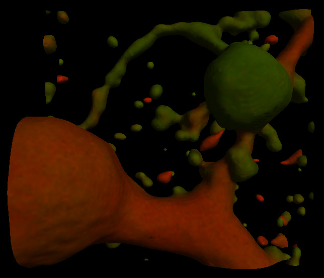
A part of the image loaded this way can be reconstructed with Smoothness Alpha=2 and Mesh Size=1 to produce the following:
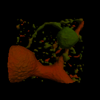
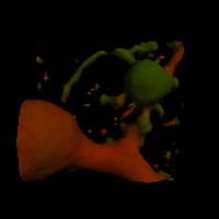
Changing the values of Smoothness and Mesh Size, we obtain the following. As Alpha becomes larger, and as Mesh Size becomes smaller, reconstruction takes more time. For Alpha=0, Mesh Size=5, it takes only a couple of seconds; but for Alpha=2, Mesh Size=1, it takes PowerBook G4 1GHz as long as 15 minutes.
| Mesh Size | Alpha=0 | Alpha=1 | Alpha=2 |
| 1 | 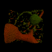 |
 |
 |
| 2 | 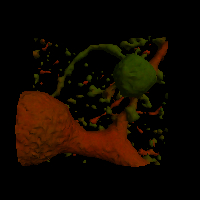 |
 |
 |
| 3 |  |
 |
 |
| 5 | 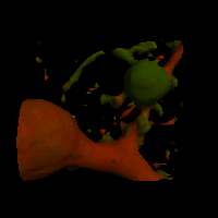 |
 |
 |